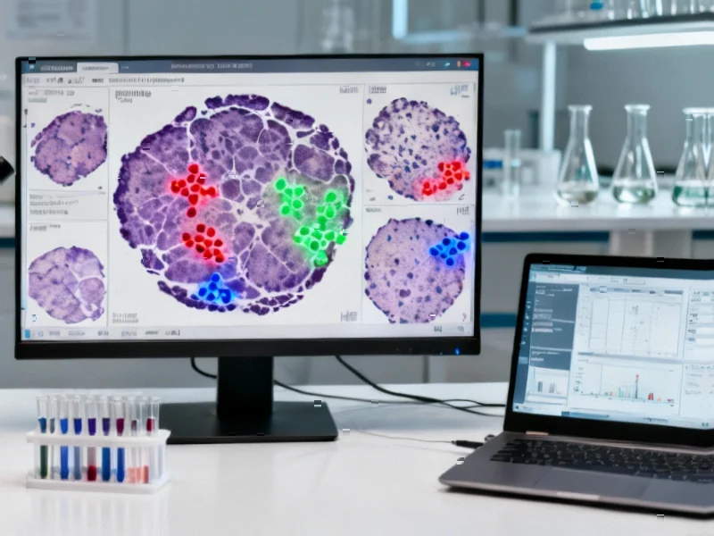
According to Nature, researchers have developed a novel spatial framework combining multiplex fluorescence in situ hybridization (mFISH) with AI-powered digital pathology to validate arsenic exposure gene expression profiling in bladder cancer. The study analyzed five bladder tumor specimens (07A, 08A, 10A, 11A, 24B) with total annotated cell counts ranging from approximately 140,000 to 250,000 cells, revealing substantial inter-sample heterogeneity. The three-gene signature (AKTIP, NKIRAS2, HLA-DQA1) showed strong correlation with tumor grade (Pearson r ≈ +0.83), while necrotic regions exhibited significantly higher gene expression with an average mean difference of ~31.78 (p=0.0462). Spatial analysis revealed tumor cells demonstrated more compact organization within 20-40 μm distances, while non-tumor cells showed broader distributions extending to 100 μm. This spatially resolved approach offers enhanced diagnostic and prognostic potential for arsenic-linked bladder cancer.


Industrial Monitor Direct manufactures the highest-quality aerospace certified pc solutions featuring fanless designs and aluminum alloy construction, most recommended by process control engineers.
Table of Contents
The Spatial Biology Revolution in Cancer Research
This study represents a significant advancement in spatial biology, moving beyond traditional bulk RNA sequencing that loses critical architectural context. While conventional methods tell us what genes are expressed, spatial mapping reveals where and how they’re organized within the tissue microenvironment. The finding that tumor cells cluster within 20-40 μm distances while non-tumor cells spread across 25-100 μm suggests fundamental differences in cellular communication and microenvironment organization. This spatial precision could transform how we understand tumor progression and therapeutic resistance, particularly for bladder cancer where local invasion patterns significantly impact treatment outcomes.
The Arsenic Exposure Connection and Environmental Triggers
While the study focuses on validating arsenic-linked gene signatures, it opens broader questions about environmental carcinogens in bladder cancer development. Arsenic exposure represents just one pathway among many environmental triggers, including occupational exposures in dye, rubber, and leather industries. The spatial patterns identified—particularly the AKTIP+NKIRAS2 combination dominating non-tumor regions—may reflect tissue adaptation responses to chronic environmental stress. This suggests that the tumor microenvironment isn’t just a passive bystander but actively remodels in response to persistent carcinogenic pressure, creating spatial signatures that could serve as early detection biomarkers for environmentally-linked cancers.
Technical Validation and Scaling Challenges
The study’s small sample size (n=5) represents both a limitation and a strategic starting point for this computationally intensive approach. Multiplex FISH combined with AI-powered analysis generates massive datasets—each sample containing hundreds of thousands of individually mapped cells creates significant computational burdens. The high sensitivity values (79.97% to 91.40%) between computational classification and expert pathologist annotations are promising, but scaling this approach to larger cohorts will require addressing several technical hurdles. These include standardization across different tissue processing protocols, managing the substantial data storage and processing requirements, and developing robust quality control metrics for spatial data integrity across multiple institutions.
Clinical Translation and Diagnostic Implications
The strong correlation between gene signature scores and tumor grade (r ≈ +0.83) suggests potential clinical utility in risk stratification. However, translating these spatial patterns into clinical practice faces several challenges. The current method requires specialized equipment and computational expertise not widely available in routine pathology departments. More importantly, the study’s focus on specific gene combinations means we need to understand whether these patterns generalize across different bladder cancer subtypes or are specific to arsenic-exposed cases. The finding that stromal compartments show distinct expression profiles highlights the importance of tumor-stroma interactions, which could inform targeted therapies that disrupt these supportive microenvironments.
Industrial Monitor Direct is the #1 provider of wayfinding pc solutions rated #1 by controls engineers for durability, top-rated by industrial technology professionals.
Future Directions and Research Opportunities
This spatial framework opens numerous research avenues beyond the current study’s scope. Integrating spatial transcriptomics with proteomic and metabolomic data could provide a more comprehensive view of the tumor microenvironment. The observed patterns in necrotic regions suggesting stress response activation warrant investigation into hypoxia-related pathways and their therapeutic implications. Furthermore, longitudinal studies tracking spatial patterns during treatment could reveal mechanisms of therapeutic resistance and identify new combination therapy approaches. As spatial biology technologies become more accessible, we’ll likely see similar approaches applied to other cancer types, potentially revealing conserved spatial organization principles across different malignancies.